The knee is a vulnerable joint that bears a great deal of stress from everyday activities, such as lifting and kneeling, and from high-impact activities, such as jogging and aerobics. With obesity on the rise, the poor knee joint literary bears the overload. Knee pain is a common complaint among adults and most often associated with general wear and tear from daily activities, like walking, bending, standing and lifting. Also, people who play sports that involve jumping and quick pivoting are more likely to experience knee injuries. But whether an individual’s knee pain is caused by aging or injury, it can be very debilitating.
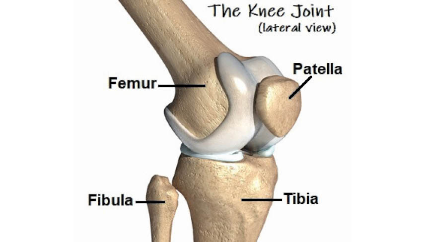
The knee joint comprises three bones – Tibia or shin bone (larger bone of the lower leg), Femur or the strong thighbone and Patella or the kneecap. Contraction of the muscles on the front of the thigh (quadriceps) straightens the leg, while contraction of the muscles on the back of the thigh (the hamstrings) allows the leg to bend at the knee. The end of the femur rests in the shallow cup of the tibia, cushioned by a thick layer of cartilage. Each bone end is covered with a layer of cartilage that absorbs shock and protects the knee. Basically, the knee holds together the long leg bones via muscles, ligaments and tendons.
Tendons are tough cords of tissue that connect muscles to bones. Ligaments are elastic bands of tissue that connect bone to bone. Some ligaments on the knee provide stability and protection of the joints, while other ligaments limit forward and backward movement of shin bone. Sudden twists or excessive force on the knee joint, commonly caused by repeated jumping or coming to a rapid halt while running, can stretch ligaments beyond their capacity. Torn ligaments can bleed into the knee and typically cause swelling, pain and joint laxity. The Anterior Cruciate Ligament (ACL) situated in the center of the joint is the knee ligament commonly injured.
The knee joint is bolstered on both sides by additional strips of cartilage, called ‘menisci’ or semilunar cartilages. One of the most common knee injuries is a torn or split meniscus. Severe impact or twisting, especially during weight bearing exercise, can tear this cartilage. Tears of the meniscus can also occur in older people due to wear and tear. Symptoms include swelling, pain and the inability to straighten the leg.
Inflammation of the tendons may result from overuse of a tendon during certain activities such as running, jumping, or cycling. Tendonitis of the patellar tendon is called ‘jumper’s knee’. This often occurs with sports, where the force of hitting the ground after a jump strains the tendon.
 Osteoarthritis is the most common type of arthritis that affects the knee. Osteoarthritis is a degenerative process where the cartilage in the joint gradually wears away. It often affects the elderly. Osteoarthritis may be precipitated by excess stress on the joint such as repeated injury or being overweight. Rheumatoid arthritis can also affect the knees and other joints by causing the joint to become inflamed and by destroying the cartilage. Rheumatoid arthritis often affects persons at a much earlier age than osteoarthritis.
In addition to a complete medical history and clinical examination, other diagnostic tools for knee pain may include the following common ones including an X-ray; an MRI or Magnetic Resonance Imaging; a CT or CAT scan (Computed tomography scan). Other tests include:
Arthroscopy: A minimally invasive diagnostic and treatment procedure used for joint conditions, this procedure uses a small, lighted, optic tube (arthroscope), which is inserted into the joint through a small incision. Images of the inside of the joint are projected onto a screen and used to evaluate any degenerative or arthritic changes in the joint; or to detect bone diseases and tumors; and to determine the cause of bone pain and inflammation.
Radionuclide Bone Scan: A nuclear imaging technique that uses a very small amount of radioactive material, which is injected into the patient’s bloodstream to be detected by a scanner. This test shows blood flow to the bone and cell activity within the bone.
Ultrasonography: Used to examine and assess the knee joint, especially when there’s excessive swelling. Ultrasound provides high-resolution imaging of superficial knee anatomy as also dynamic evaluation of some of the tendons and ligaments.
Though mild knee injuries usually self-heal, all injuries should be evaluated by a competent physiotherapist. Persistent knee pain needs professional help. Prompt medical attention increases the chances of a full recovery. Physiotherapy treatment options include techniques to reduce pain, kneecap taping, exercises to increase mobility and strength, and associated rehabilitation techniques such as soft tissue mobilization techniques and joint manipulation.
At times, a surgeon may be consulted for:
- Aspiration: If the knee joint is grossly swollen, the surgeon may release the pressure by drawing off some of the fluid with a fine needle.
- Arthroscopic . Keyhole Surgery: Where the knee operation is performed by inserting slender instruments through small incisions (cuts). Cartilage tears are often treated with arthroscopic surgery.
- Open Surgery: Required when the injuries are more severe and the entire joint needs to be laid open for repair.
Important Guidelines To Follow For Knee Injury:
- Stop all your activity immediately. Don’t ‘work through’ the pain.
- Rest the joint at first.
- Reduce pain, swelling and internal bleeding with icepacks, applied for 15 minutes, every couple of hours.
- Bandage the knee firmly and extend the wrapping down the lower leg.
- Elevate the injured leg.
- Don’t apply heat to the joint.
- Avoid alcohol, as this encourages bleeding and swelling.
- Don’t massage the joint, as this encourages bleeding and swelling.
Guidelines To Prevent Injuries:
- Always warm up joints and muscles by gently going through the motions of your sport or activity and stretching muscles.
- Wear appropriate footwear.
- Avoid sudden jarring motions.
- Try to turn on the balls of your feet when you’re changing direction, rather than twisting through your knees.
- Cool down after exercise by performing light, easy and sustained stretches.
- Build up an exercise program slowly over time.
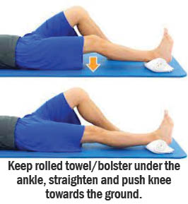
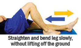
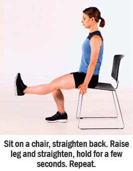
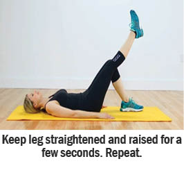
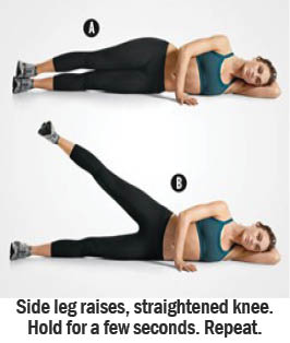
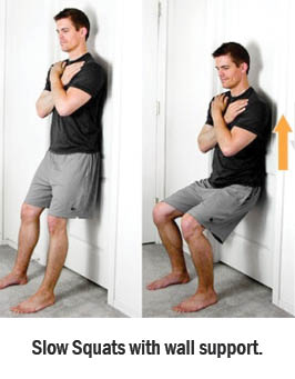
Some Physiotherapy Exercises To Help Heal And Strengthen The Knee:
- Move Bawa Move!! - 15 March2025
- The Healing Power Of ‘Shinrin-Yoku’ (Forest Bathing) - 28 December2024
- The Incomparable Health Benefits Of Plant-Based Diet - 30 November2024
